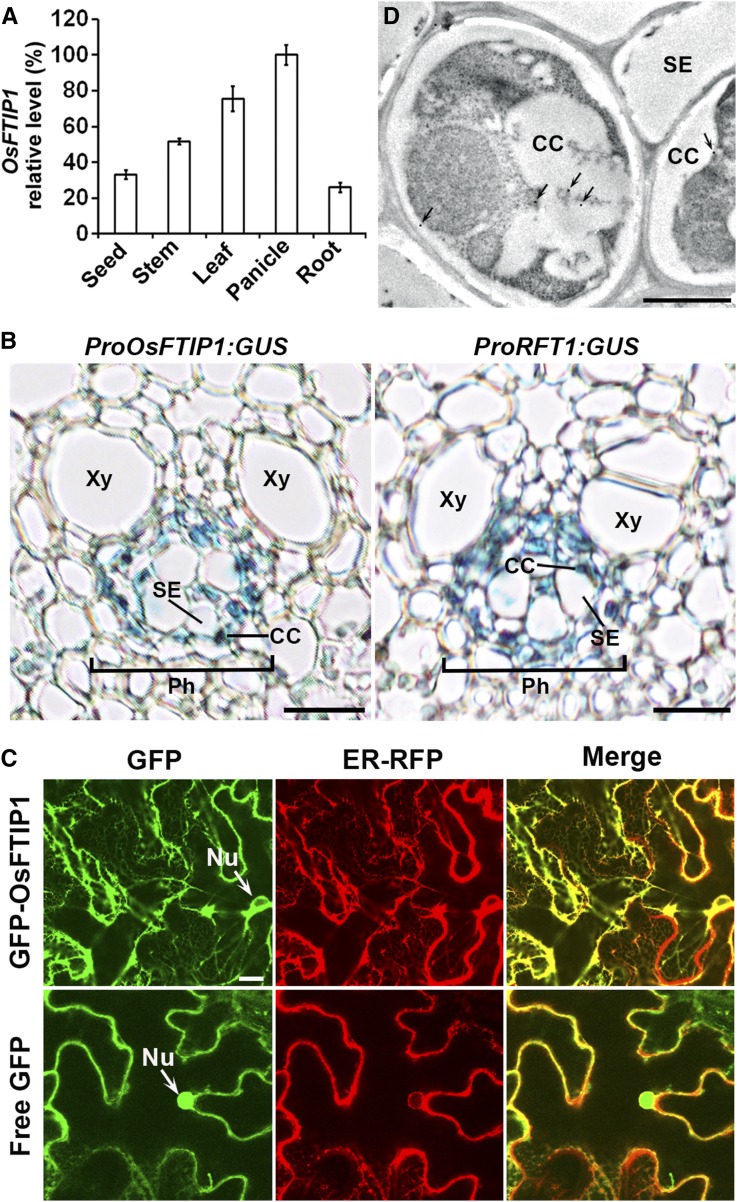
Figure 2.
Expression Patterns of OsFTIP1.
(A) Quantitative real-time PCR analysis of OsFTIP1 expression in various tissues of wild-type plants grown under LDs. Results were normalized against the expression levels of Ubiquitin based on three biological replicates. Error bars indicate sd.
(B) Transverse section of the leaf blade from representative ProOsFTIP1:GUS and ProRFT1:GUS transgenic plants grown under LDs at 50 DAS. CC, companion cell; Ph, phloem; SE, sieve element; Xy, xylem. Bars = 10 μm.
(C) Subcellular localization of GFP-OsFTIP1and free GFP in N. benthamiana leaf epidermal cells. GFP-OsFTIP1 is mostly colocalized with an ER marker. GFP, GFP fluorescence; ER-RFP, RFP fluorescence of an ER marker; Merge, merge of GFP and RFP; Nu, nucleus. Bar = 10 µm.
(D) Analysis of OsFTIP1-HA localization by immunogold electron microscopy using anti-HA antibody in companion cell-sieve element complexes in the leaf vasculature of Osftip1-1 gOsFTIP1-HA. Arrows indicate the locations of gold particles. CC, companion cell; SE, sieve element. Bar = 1 µm.

