Fig. 2.

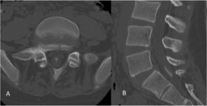
Plain computed tomography (CT) at L5/S1 level (A) and reconstructive CT images at the L5–S2 level (B) showed an ossified lesion within the dura mater.

Plain computed tomography (CT) at L5/S1 level (A) and reconstructive CT images at the L5–S2 level (B) showed an ossified lesion within the dura mater.