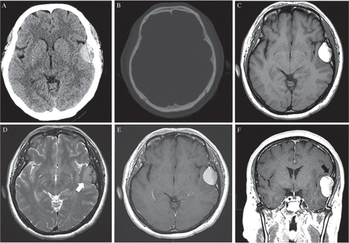
Fig. 1.
(A) CT imaging revealed a mass (diameter: 3.2 cm) in the left temporal convexity. (B) Bone imaging of CT revealed no skull fracture. (C) Axial T1-weighted MR imaging revealed a convex, well-defined mass with homogenous high signal intensity. (D) Axial T2-weighted MR imaging revealed a cerebrospinal fluid (CSF) cleft (white arrow) along the medial boundary. (E) The mass did not show significant enhancement. (F) Coronal, post-gadolinium, T1-weighted MR imaging revealed the dural tail sign (black arrow) at the superior portion of the lesion.

