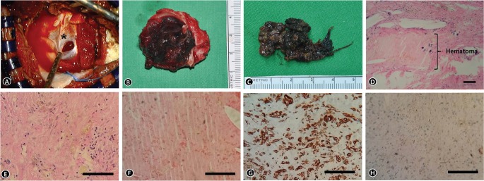
Fig. 2.
(A) Intraoperative photograph showing the meningeal layers (asterisk) of the dura mater after the craniotomy. (B) The hematoma was removed with the periosteal layer of the dura matter because this was adhered to the bone flap. (C) The hematoma was an encapsulated, fibrous, dark-colored, bloody mass. (D) Microphotography of the dura matter revealed a fibrocollagenous tissue (periosteal layer of dura matter and meningeal layer of dura matter) with interdural hematoma (H&E, ×40). (E) Microphotography of the dura matter revealed an eosinophilic lining and scattered inflammatory cells in the lesion (H&E, ×100). (F) Magnified microphotography of the hematoma (H&E, ×100). (G) Immunohistochemical staining for epithelial membrane antigen (EMA) revealed negative in the specimen (×100). (H) Immunohistochemical staining for Vimentin revealed positive in the specimen (×100).

