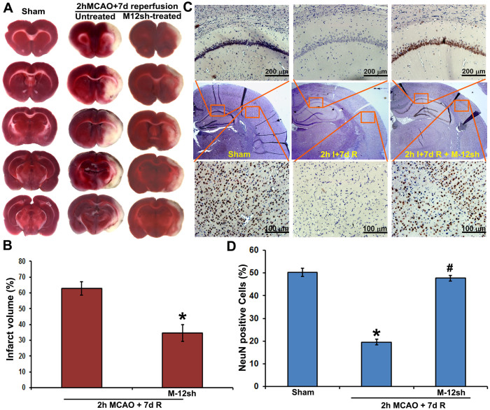
Figure 5. In vivo efficacy of plasmids expressing MMP-12 shRNA.
(A) Representative TTC staining images of the rat coronal brain sections of sham-operated, untreated MCAO-subjected and MMP-12 shRNA (M-12sh) treated MCAO-subjected rats sacrificed seven days after reperfusion. n = 6. The white-colored areas represent the infarct regions in these sections, and the red-colored areas represent normal areas. (B) Quantification of infarct volume from TTC stained sections using image analysis software. The possible influence of edema on infarct volume was corrected by standard methods (volume of contralateral hemisphere − volume of non-ischemic ipsilateral hemisphere), with infarcted volume expressed as a percentage of the contralateral hemisphere. n = 6. Values are expressed as mean ± SEM; *p < 0.05 vs. untreated, MCAO-subjected animals. (C) Immunohistochemical analysis of ipsilateral rat coronal brain sections depicting DAB staining (brown), representative of neurons. The missing NeuN-DAB staining in injured brain sections due to neuronal loss is restored after MMP-12 knockdown. Brain sections are counterstained with hematoxylin for nuclear localization. Results are from six independent sections obtained from six different rats. I-ischemia; R-reperfusion. (D) Quantification of NeN positive cells in the cortex of ipsilateral brain. n = 6. *p < 0.05 vs. sham; #p < 0.05 vs. untreated MCAO-subjected animals.

