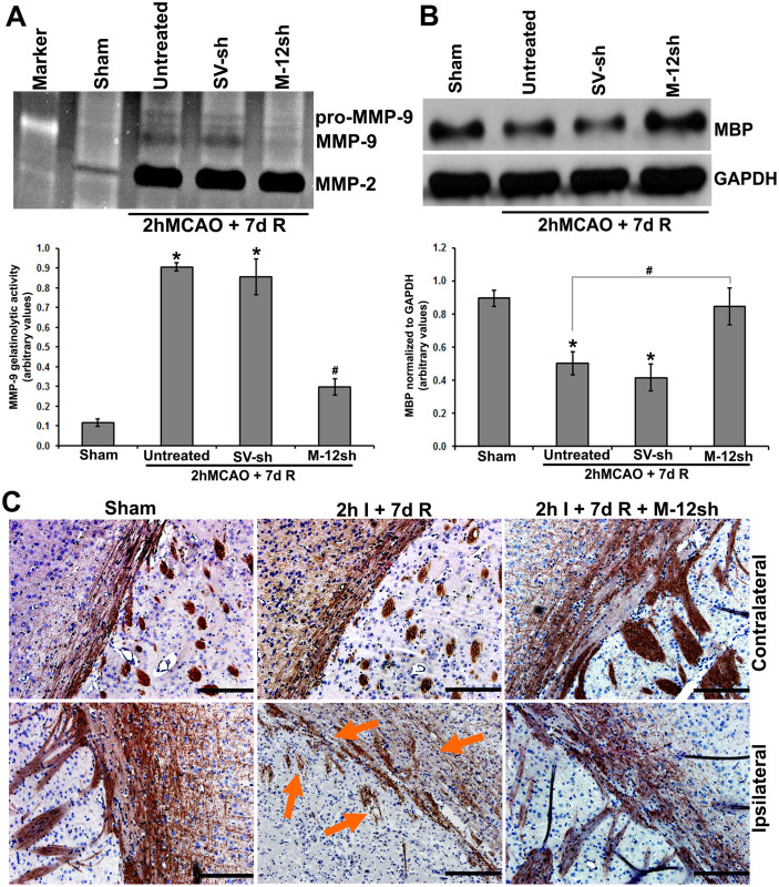
Figure 6. Effect of MMP-12 knockdown on other possible MMP-12 substrates.
(A) Gelatin zymogram showing MMP levels in sham control and ischemic brains of rats subjected to a two-hour MCAO followed by seven days reperfusion without treatment and treatment with plasmids containing a vector ligated with a scrambled sequence (SV-sh) and a vector ligated with MMP-12 shRNA (M-12-sh). Quantification of MMP-9 gelatinolytic activity. n = 6. *p < 0.05 vs. sham; #p < 0.05 vs. untreated. R = reperfusion (B) Immunoblot showing the protein expression of MBP in the ischemic brains of rats subjected to a two-hour MCAO followed by seven days reperfusion with and without treatments. GAPDH was used as a loading control. Quantification of MBP protein expression. n = 6. *p < 0.05 vs. sham; #p < 0.05 vs. untreated. (C) DAB immuno-staining depicting the protein expression of MBP in the contralateral and ipsilateral rat coronal brain sections obtained from various groups of rats. Arrows demonstrate the marked loss of MBP-immunostained axonal processes with clear structural abnormalities of rarefaction and fragmentation. Brain sections are counterstained with hematoxylin for nuclear localization. n = 6. Scale bar = 200 μm.

