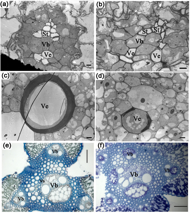
Figure 8. Disturbed vasculatures in the osnpf2.2-1 mutants, as shown by transmission electron microscopy and semisection analysis.
(a, b) Anther cross section from a wild-type (WT) plant (a) and an osnpf2.2-1 mutant (b); the sieve tubes (St) and vessels (Ve) in the osnpf2.2-1 mutant are disrupted; bar = 2 µm. (c, d) Ultrastructure of leaf blades from a WT plant (c) and an osnpf2.2-1 mutant (d); a few vessels in the venation in the mutant plant showed delayed dying as compared with those in the WT plant; bar = 5 µm. (e, f) Semisection of primary branches from a WT plant (e) and an osnpf2.2-1 mutant (f), bar = 50 µm. There was no obvious vascular sheath around the vascular bundle (Vb) in the osnpf2.2-1 mutant, and its sieve tubes and vessels were irregularly distributed in the vascular bundles.

