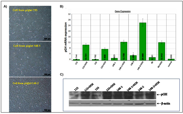
Figure 5. Expression of pGH in cells from F0 transgenic pigs in the presence and absence of DOX.
A) Ear-tip fibroblasts were separated from partial transgenic pigs. B) QRT-PCR analysis of pGH mRNA expression in ear-tip fibroblast cells from transgenic pigs. C) Western blot analysis of pGH protein detection in cells from transgenic pigs. After DOX induction, pGH protein levels increased significantly compared with those in the control and the non-induced groups. The blots have been cropped to focus on the bands of interest. See Supplementary Fig. S6 for full-length gels.

