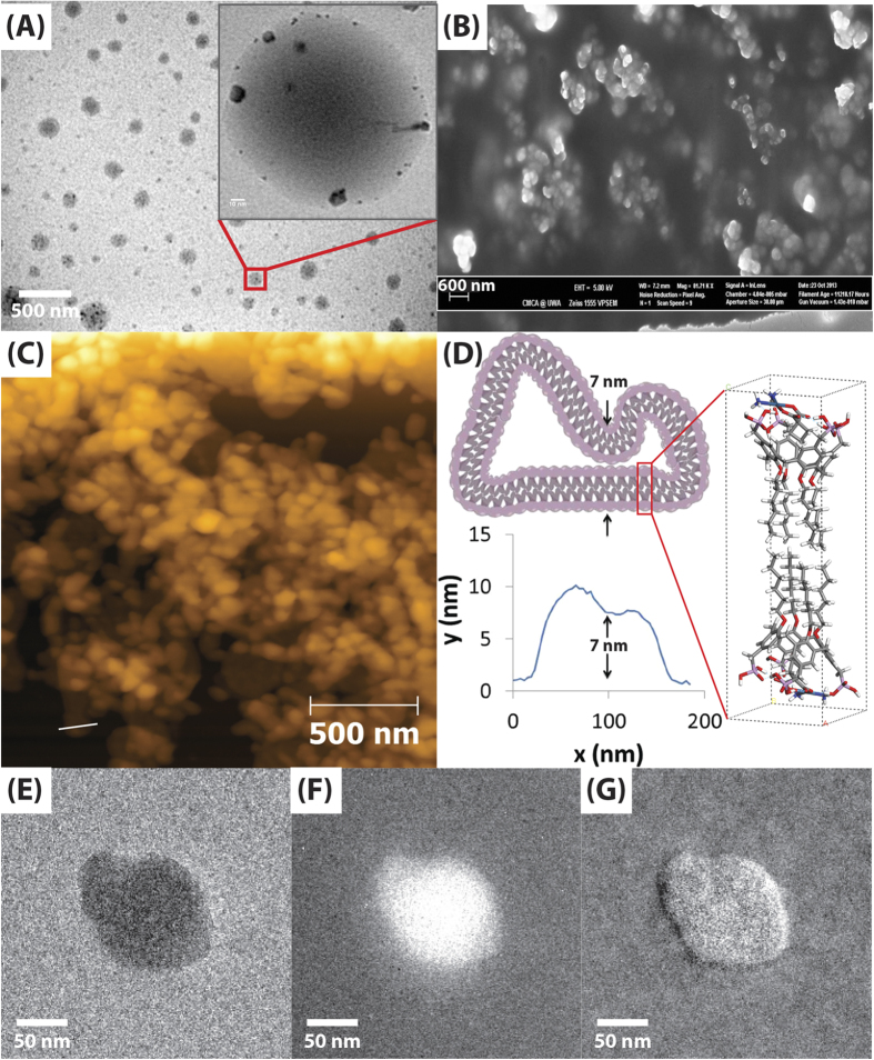
Figure 1.
VFD processed P4C6-carboplatin host-guest vesicles: (A) TEM image, scale bar 500 nm (10 nm for inset), (B) SEM image, scale bar 600 nm, (C) AFM image, (D) sectional height profile of a collapsed vesicle and elemental mapping of host-guest complex with energy-filtered transmission electron microscopy (EFTEM) for (E) unfiltered, (F) carbon and (G) platinum.

