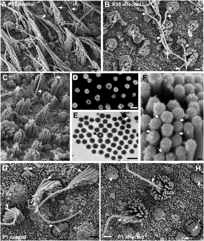
Fig 7. Utricular macula.
A. Normal. Aged P35. Stereociliary bundles are elongated in shape. Well-defined gradients in the height of stereocilia across the bundle. The longest stereocilia adjacent to the kinocilium (arrowed) are very long. B-F Affected animals. B. P35 littermate of the normal animal shown in A. Bundles and apical surface rounded. Stereocilia appear shorter than normal. Long kinocilia (arrowed) emerge from the centre of some bundles, but in other bundles the kinocilium does not rise above the stereocilia. C. P42. A few bundles show distinct height gradients from periphery to centre of the bundle, but in the majority the height gradients are not marked or stereocilia are of similar height, Arrows indicate kinocilium emerging from the centre of stsreociliary bundles. D. Phalloidin labelling of actin in stereocilia in a whole mount preparation, shows every bundle in the field is affected, each bundle is round with the stereocilia surrounding a central unlabelled “hole”. E. Thin section across a hair bundle. Stereocilia surround a centrally located kinocilium (arrowed). F. Tip-link like structures (arrows) from tip of shorter stereocilium to side of adjacent longer oriented from periphery to centre. G-H Maturation of hair bundles. G. Normal at P1. Progressive stages in the maturation of the hair bundles are evident. In all bundles from the most immature with the shortest stereocilia, kinoclia (arrowed) are positioned eccentrically behind the longest stsreocilia. H. Affected animal at P1. In all bundles, from the most immature, kinocilia (arrowed) are located in the centre of the cell and are completely encircled by stereocilia which cover the entire apical surface of the cell. Scale bars: A,B,C 1μm; D 10μm; E 0.5μm; F 100nm; G, H,I 1μm.

