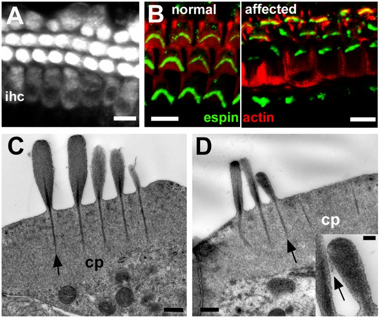
Fig 9. Defects are confined to hair bundle organisation.
A. FM1-43 uptake in whole mount preparation of the organ of Corti of an affected animal at P10. After brief exposure to FM1-43, dye enters both outer hair cells and inner hair cells (ihc). Scale bar: 10μm. B. Immuno-labelling for espin (green) in whole mount preparation of the organ of Corti (actin labelled with rhodamine tagged phalloidin, red) of a normal and an affected animal at P22. Stereocilia in morphologically abnormal bundles label as intensely for espin as those in the normal littermate. Scale bar: 10μm. C, D. Thin sections of the apical end of inner (C) and outer (D) hair cells in affected animal at P30. Cuticular plates (cp) are well developed. Prominent stereociliary rootlets (arrows) descend from the shafts of the stereocilia into cuticular plates. Lateral cross-links between shafts of stereocilia arrowed in inset. The longest stereocilia in the bundle of the IHC (C) are quite short and there are two “long” stereocilia in adjacent rows of the same height. In the OHC (D) the two longer stereocilia in adjacent rows are of similar height while the shorter stereocilium appears unusually wide. In the inset the lateral crosslinking between adjacent stereocilia is indicated by the arrow. Scale bars: A,B 10μm; C, D 0.5μm, inset 0.1μm.

