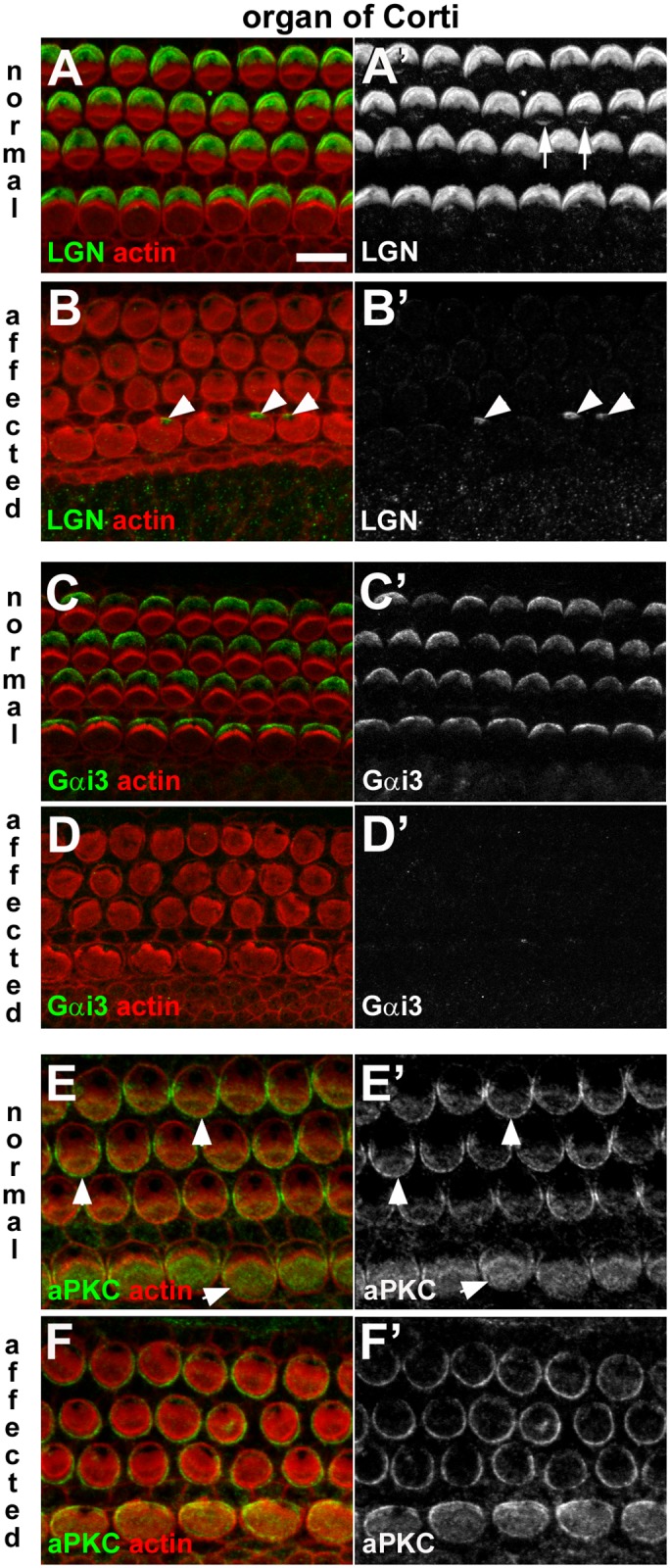
Fig 11. Dysregulation of apical surface asymmetry in cochlear hair cells of affected animals.

A-B: LGN in normal and affected animals. In a whole-mount of P1 normal organ of Corti the anti-LGN antibody labelled the surface of OHC and IHC, specifically at the lateral pole (A, A’). Labelling at the tips of stereocilia also evident (arrows in A’). The asymmetric pattern of LGN distribution was not apparent in an affected littermate (B, B’). LGN immunofluorescence was detected in the peri-centriolar region of some IHC of affected animals (arrowheads in B’). C-D: Gαi3 in normal and affected animals. Gαi3 at the surface of hair cells in the normal organ of Corti (C,C’) showed the same asymmetric distribution as LGN. In affected animals labelling for Gαi3 is virtually undetectable in the organ of Corti (D,D’). E-F: aPKC in normal and affected animals. aPKC was detected at the surface of normal hair cells in the organ of Corti (E,E’), most strongly on the medial side (i.e. the side opposite to that of LGN and Gαi3 labelling) (arrowheads). In an affected littermate aPKC was detected in hair cells in the organ of Corti (F,F’), but the asymmetric distribution was not apparent. Scale bar in A (refers to all figures): 10 μm.
