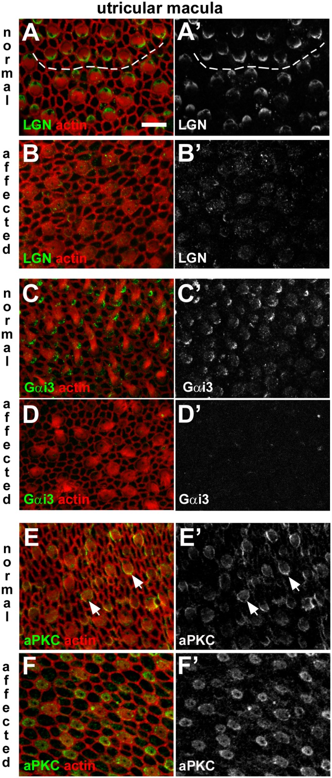
Fig 12. Dysregulation of apical surface asymmetry in utricular hair cells of affected animals.

A-B: LGN in normal and affected animals. In a whole-mount of P1 normal utricular macula the anti-LGN antibody labelled the surface of hair cells, specifically at one side (A, A’). The micrograph details the striolar region in which hair cells of opposing orientation are apparent. The dashed line traces the line of polarity reversal. The asymmetric pattern of LGN immunofluorescence was not apparent in an affected littermate (B, B’). C-D: Gαi3 in normal and affected animals. Immuno-labelling for Gαi3 at the surface of hair cells in the normal utricular macula (C,C’) shows the same asymmetric distribution as LGN. In affected animals labelling for Gαi3 is virtually undetectable (D,D’). E-F: aPKC in normal and affected animals. aPKC was detected at the surface of hair cells in the normal utricular macula (E,E’), most strongly on the medial side (i.e. the side opposite to that of LGN and Gαi3 labelling; arrows). In an affected littermate aPKC was detected in hair cells (F,F’), but the asymmetric distribution was not apparent. Scale bar in A (refers to all figures): 10 μm.
