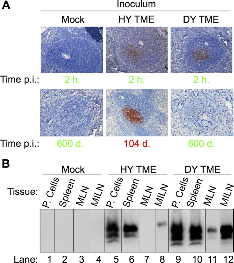
Fig 3. Rapid transport of HY and DY PrPSc to lymphoid tissues following intraperitoneal inoculation.
Hamsters (n = 3 per inoculum) were intraperitoneally inoculated with brain homogenate from uninfected (Mock), HY TME or DY TME-infected hamsters and PrPSc was detected with either A) immunohistochemistry or B) Western blot. A) Spleen from negative control mock inoculated animals did not contain detectable PrPSc at 2 hours p.i. or at 600 d. p.i.. Spleen from positive control HY TME infected hamsters contained PrPSc immunoreactivity in the germinal center of lymphoid follicles at both 2 hours p.i. and during the clinical phase of disease. PrPSc immunoreactivity was detected in spleen from DY TME infected hamsters at 2 hours p.i. but not at 600 days p.i.. Incubation periods in green text are clinically normal and in red text are clinically affected. B) Western blot of peritoneal cells (P. Cells), spleen, mesenteric lymph node (MLN) and medial iliac lymph node (MILN) from hamsters intraperitonally inoculated with uninfected (Mock), HY TME or DY TME-infected brain homogenate at 2 hours p.i.

