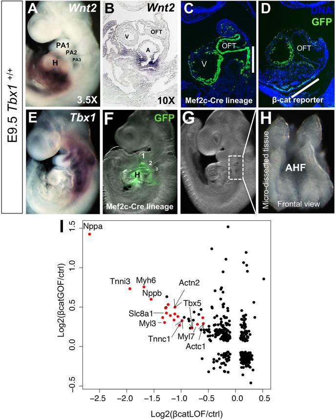
Fig 1. Constitutive β-catenin expression in the AHF promotes differentiation.
(A) Whole mount in situ hybridization (WMISH) of Wnt2 in a wild type (Tbx1+/+) embryo at embryonic day 9.5 (E9.5) and a sagittal section from the heart region of the embryo is shown in B. (C) Mef2c-AHF-Cre lineage in a wild type embryo in a sagittal section. (D) Visualization of β-catenin signaling using a TCF/Lef:H2B-GFP reporter allele in a sagittal section of a wild type embryo. (E) WMISH of Tbx1 in a wild type embryo. (F) Mef2c-AHF-Cre lineage shown by green fluorescence in a wild type embryo (Mef2c-AHF-Cre/+;ROSA26-GFP f/+) at E9.5. Numbers denote the pharyngeal arches 1, 2 and 3. (G) Whole mount embryo showing the region that has been dissected for the experiments (rectangle in dorsal to the heart). (H) The micro-dissected region containing the Mef2c-AHF-Cre lineage is shown from a frontal view. (I) Comparison of global gene expression changes in micro-dissected tissues of β-catenin GOF and β-catenin LOF embryos at E9.5. Differentially expressed genes (p < 0.05 and FC > 1.5) in at least one of the two comparisons, β-catenin LOF vs control (x-axis) or β-catenin GOF vs control (y-axis), were plotted. Red dots denote cardiac differentiation genes. Abbreviations: heart (H), pharyngeal arch (PA), outflow tract (OFT), ventricle (V), atrium (A), FC = fold change.

