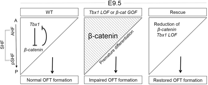
Fig 7. Model for Tbx1 and β-catenin function in the SHF.
In the model, the triangle represents the Mef2c-AHF-Cre lineage and the Tbx1 expression pattern, before migrating into the heart tube at E9.5. Left panel: Tbx1 expression is strongest in the AHF and weakest in the posterior SHF (pSHF) while Wnt/β-catenin expression is opposite. Left panel depicts a possible double negative feedback loop between the two genes in the SHF, required for normal cardiac outflow tract (OFT) formation. Middle panel shows increased differentiation in the AHF when Tbx1 is inactivated or β-catenin is constitutively active in the AHF. This results in premature differentiation within this tissue. Right panel depicts the rescue genotype in which both alleles of Tbx1 and one allele of β-catenin was inactivated in the AHF. Significant rescue of heart defects was obtained. Abbreviations: A = anterior, P = posterior, OFT = outflow tract.

