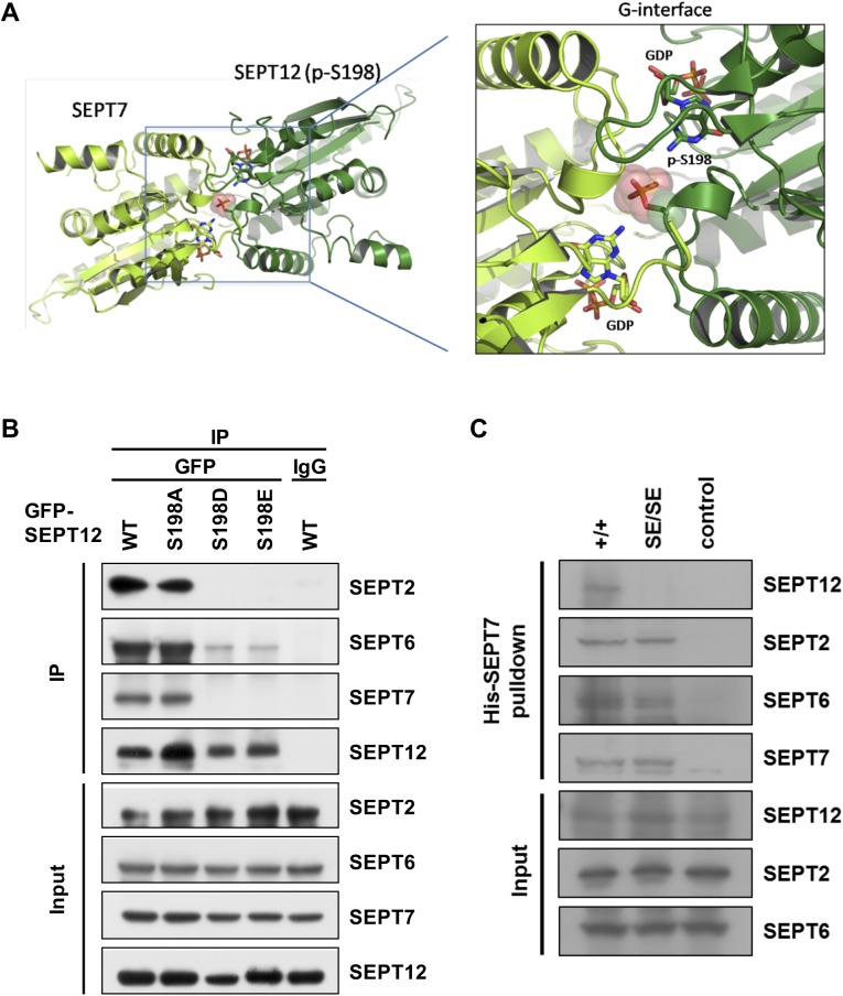
Fig 5. Mimetic phosphorylated Ser198 of SEPT12 disrupts SEPT12-7-6-2 complex.
(A) The 3D-structure of the SEPT12-SEPT7 complex model shows that phosphorylated Ser198 (p-S198) of SEPT12 (shown in pink) is located at the dimeric G-interface. The structure of SEPT12 was built based on SEPT7 (PDB code: 3T5D) using the homology modeling method on the Swiss-model server (http://swissmodel.expasy.org/). (B) Myc-SEPT6 and FLAG-SEPT7 were co-transfected with various GFP-SEPT12 plasmids into NT2/D1 cells, and the cell lysates were immunoprecipitated using an anti-GFP antibody. The expression of SEPT2, 6, 7 and SEPT12 was detected using anti-SEPT2, anti-Myc, anti-FLAG and anti-GFP antibodies, respectively. (C) His-SEPT7 pull-down assay in WT and SEPT12 KI testis. His-SEPT7 recombinant protein was incubated with Ni-NTA beads followed by testicular lysate from WT and SEPT12S196E/S196E mice. The pull-down protein was collected and subjected to western blotting for SEPT12, SEPT2 and SEPT6. The level of His-SEPT7 was detected using anti-His antibody. For the control group, WT testicular lysate was incubated with BSA instead of His-SEPT7.

