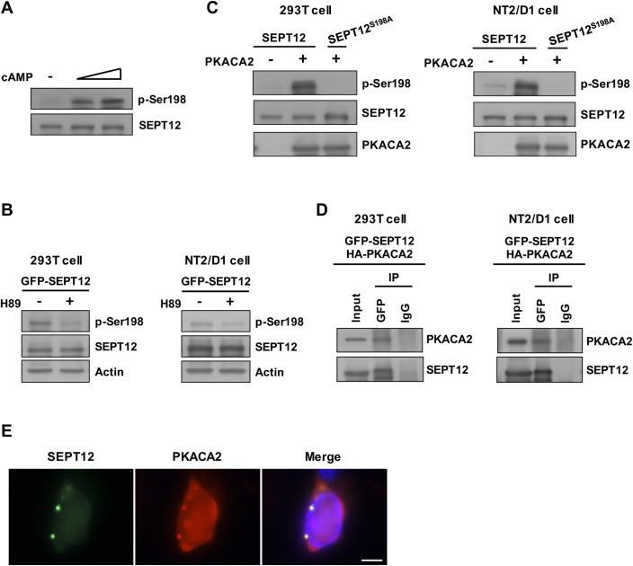
Fig 6. The SEPT12 Ser198 residue was phosphorylated by PKA.
(A) The Ser198 phosphorylation of SEPT12 (p-Ser198) was gradually elevated through increasing the dosage of cAMP treatment. GFP-SEPT12 was overexpressed in 293T cells, followed by treatment with 0, 1, and 5 mM cAMP, and the expression of phosphorylated Ser198 and SEPT12 was detected using anti-phospho-Ser198 and anti-GFP antibodies, respectively. (B) GFP-SEPT12 was overexpressed in 293T and NT2/D1 cells, followed by treatment with 50 μM H89, and the expression of phosphorylated Ser198, SEPT12 and actin was detected using anti-phospho-Ser198, anti-GFP and anti-Actin antibodies, respectively. (C) GFP-SEPT12 and GFP-SEPT12S198A, with or without HA-PKACA2, were transfected into 293T cells and NT2/D1 cells as indicated, and the expression of phosphorylated Ser198, SEPT12 and PKACA2 was detected using anti-phospho-Ser198, anti-GFP and anti-HA antibodies, respectively. (D) Co-immunoprecipitation of GFP-SEPT12 with HA-PKACA2. 293T Cells or NT2/D1 cells were transfected with GFP-SEPT12 and HA-PKACA2, and the lysates were immunoprecipitated using an anti-GFP antibody. Immunoblotting was performed using anti-GFP and anti-HA antibodies. (E) SEPT12 and PKACA2 showed partial co-localization. GFP-SEPT12 and HA-PKACA2 were co-expressed in NT2/D1 cells, and immunofluorescence was detected using an anti-HA antibody. Scale bar, 10 μm.

