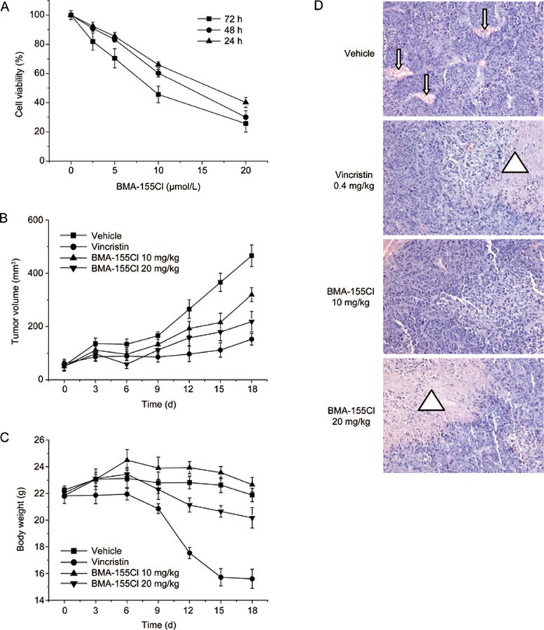
Figure 2.
BMA-155Cl's antitumor effect in vitro and in vivo. (A) HepG-2 cells were treated with 2.5–20 μmol/L BMA-155Cl respectively for 24–72 h. Cells viability was estimated by MTT assay. Data were obtained from three independent experiments and presented as the mean±SD. BMA-155Cl inhibited the growth of tumor volume (B) and body weight (C) in vivo. Data were presented as mean±SD. n=6. (D) BMA-155Cl induced the necrosis of the tumor tissue after H&E staining. The blood vessels were presented as red regulated pattern in vehicle group (pointed by the arrow), whereas the necrosis zones were presented as patches of red in vincristine and BMA-155Cl groups (pointed by the triangle). Magnification ×200. *P<0.05, **P<0.01 vs vehicle.

