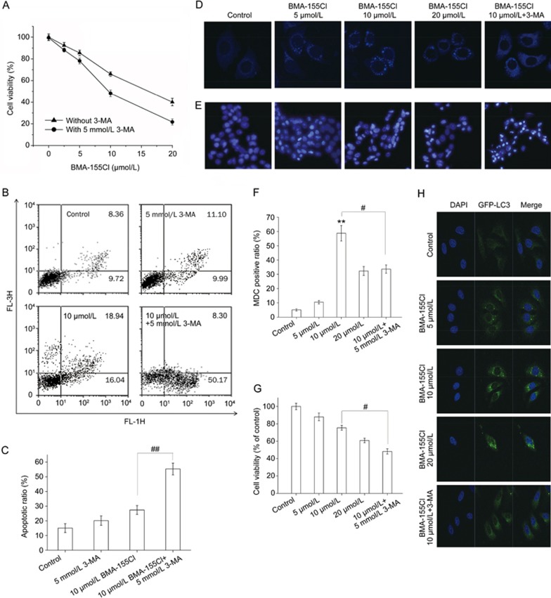
Figure 5.
Inhibition of autophagy by 3-MA potentiates BMA-155Cl's pro-apoptotic effect. HepG-2 cells were treated with various concentrations of BMA-155Cl (5–20 μmol/L) for 24 h in the absence or presence of 3-MA (5 mmol/L, pretreated for 1 h). The cell viability ratio (A) and the apoptotic ratio (B) were measured by MTT and flow cytometry. The MDC staining (D) and DAPI staining (E) assay were performed by confocal microscopy. The corresponding quantified histograms of B, D and E are presented as C, F and G, respectively. (H) BMA-155Cl induced accumulation of GFP-LC3 puncta in U87 cells in a dose-dependent manner within 24 h. Magnification ×40. All data were expressed as the mean±SD of triplicate experiments. #P<0.05, ##P<0.01 vs BMA-155Cl 10 μmol/L.

