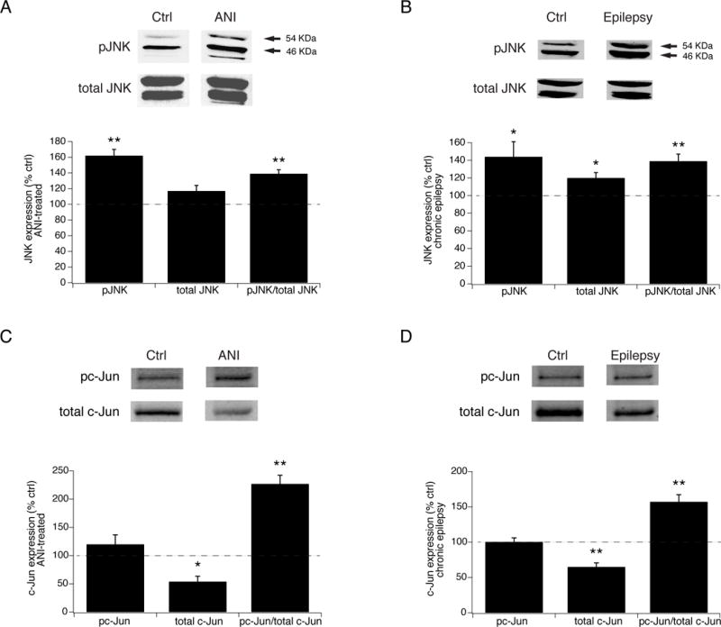
Fig. 7.

JNK signaling is hyperactivated in chronic epilepsy. A. Summary data showing increased JNK activation in hippocampal tissue from naive animals after in vitro treatment with ANI compared to untreated tissue. Shown are elevated phospho-JNK (pJNK) and phospho- to total JNK levels in ANI-exposed tissue, while total JNK levels remained unchanged. Above are representative blots against pJNK and total JNK in ANI-exposed and naive controls. Arrows denote 54 and 46 kDa bands. B. In hippocampal tissue from chronically epileptic animals, pJNK levels are increased compared to those from naive, non-epileptic controls, along with modestly activated total JNK levels, and an increased ratio of phospho- to total JNK expression. C. In vitro ANI treatment of hippocampal tissue from naive animals produced a significant increase in the fraction of phosphorylated c-Jun, while producing a decrease in the level of total c-Jun expression compared to untreated tissue. D. Phosphorylated c-Jun was increased in tissue from chronically epileptic animals compared to that in naive, nonepileptic animals, and similarly to ANI-exposed tissue, demonstrated a decrease in total c-Jun levels.
