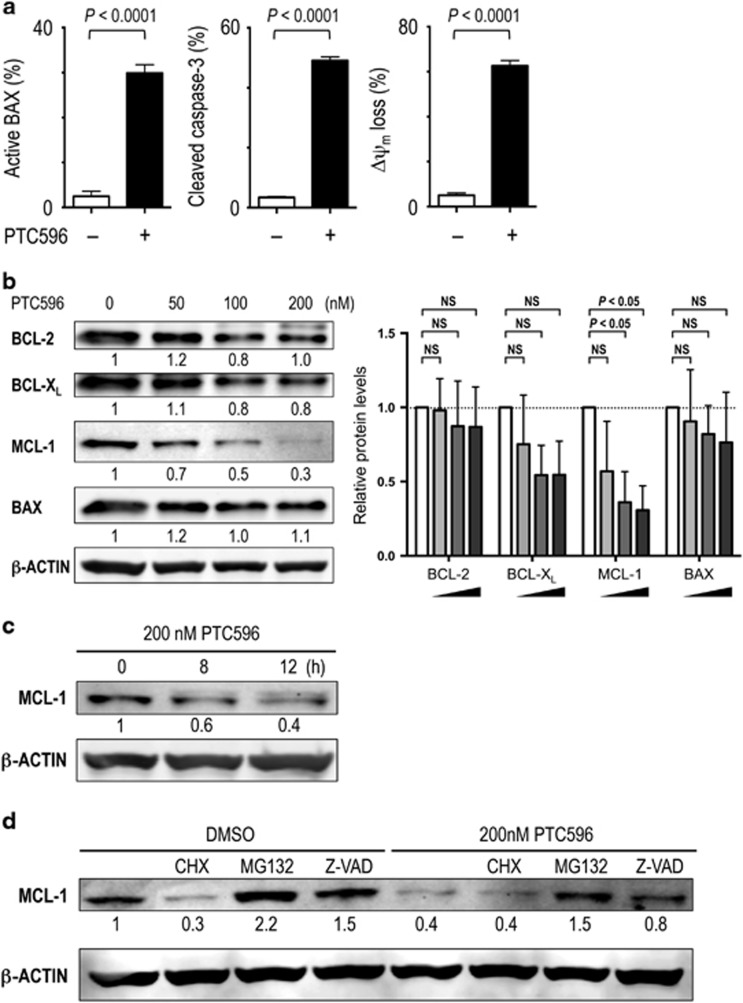
Figure 4.
PTC596 reduces anti-apoptotic MCL-1 levels and induces mitochondrial apoptosis in AML. (a) BAX conformational changes, caspase-3 cleavage and Δψm loss were determined by flow cytometry in MOLM-13 cells after 16-h (BAX activation) or 20 h (caspase-3 cleavage and Δψm loss) exposure to 200 nM PTC596. The results are expressed as the mean±s.d. (b) Expression of BCL-2 family proteins in MOLM-13 cells after 20-h treatment with 50, 100 or 200 nM PTC596. The intensities of immunoblot signals were quantified and normalized to those of β-ACTIN. Levels in untreated cells were set at 1.0. White bars represent untreated controls. One-way ANOVA with Dunnett's test was used to determine significance between control and treated samples. NS, not significant. (c) MCL-1 expression in MOLM-13 cells treated with 200 nM PTC596 for indicated time periods. (d) MOLM-13 cells were preincubated for 1 h with 7 μM cycloheximide (CHX), 0.5 μM MG132 or 25 μM Z-VAD-FMK, and MCL-1 levels were determined after 20-h treatment with 200 nM PTC596. β-ACTIN was used as loading control. Results are representative of three independent experiments.

