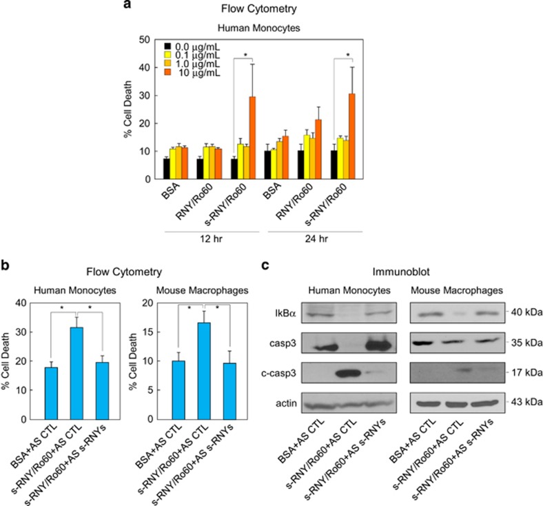
Figure 3.
Extracellular s-RNYs promote apoptosis and inflammation in lipid-laden macrophages. (a) Time and dose response of cell death by flow cytometry from human THP1 cells incubated with the immunopurified complex of s-RNY/Ro60 or RNY/Ro60. Incubation with BSA was used as control. Data are presented as mean and S.D. (n=5). One-way ANOVA with post-hoc Tukey test: *P<0.05. (b) Flow cytometry from human THP1 cells (left panel) or mouse BMDMs (right panel) incubated with 10 μg/ml of either the immunopurified complex of s-RNY/Ro60 or BSA, as control. s-RNY/Ro60 complex was previously incubated for 16 h in rotation at 4 °C with either 40 nM of 2'-OMe-RNA AS to s-RNYs or control. Data are presented as mean and S.D. (n=5). Student's t-test: *P<0.05. (c) Immunoblot analysis of IκBα, actin, total caspase 3 (casp3) and its cleaved form (c-casp3) in THP1 cells (left panel) or BMDMs (right panel) incubated s-RNY/Ro60 complex previously pretreated with either AS to s-RNYs or control

