Figure 5.
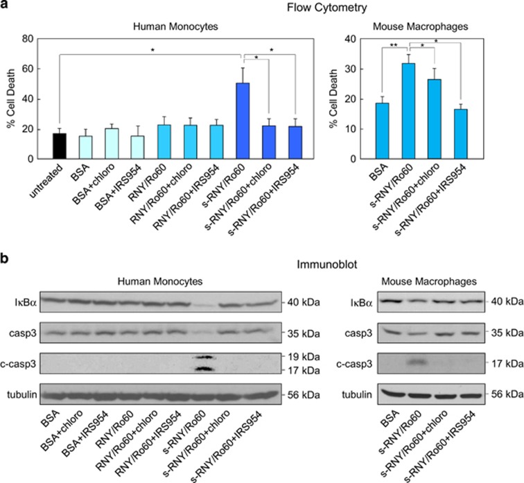
Extracellular s-RNYs activate TLR7 to promote apoptosis and inflammation in monocytes/macrophages. Cell death data by flow cytometry from human THP1 cells (a – left panel) or mouse BMDMs (a – right panel) incubated with 10 μg/ml of the immunopurified complex of s-RNY/Ro60, RNY/Ro60 or BSA. Cells were incubated with 50 mM of chloroquine for 24 h or with IRS954 at 10 μg/ml of concentration for 24 h, as indicated. Percentage of apoptotic cells was determined by staining with annexin V-FITC. Data are presented as mean and S.D. (n=6 for BMDMs and 4 for THP1 cells). The same experimental conditions were analyzed by immunoblot of IκBα, actin, total caspase 3 (casp3) and its cleaved form (c-casp3) in THP1 cells (b – left panel) or BMDMs (b – right panel). Student's t-test: *P<0.05; **P<0.01

