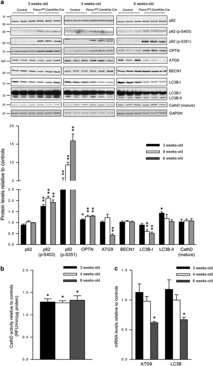
Figure 4.
26S proteasome dysfunction induces selective autophagy, but continued dysfunction decreases essential autophagy proteins. (a) Representative immunoblots and quantification at 3, 4 and 6 weeks old of control and Psmc1fl/fl;CaMKIIα-Cre cortices. Short and long exposures for LC3B are shown. Glyceraldehyde 3-phosphate dehydrogenase (GAPDH) was used as a loading control at each age for quantification; a representative GAPDH is shown. Error bars represent S.E.M. of n≥3 mice. *P<0.05 and **P<0.01 by unpaired Student's t-test. (b) CathD activity in 3, 4 and 6-week-old control and Psmc1fl/fl;CaMKIIα-Cre cortices. Pepstatin A was used as a control and decreased CathD activity to zero values (data not shown). Error bars represent S.E.M. of n=5 mice. *P<0.05 by unpaired Student's t-test. (c) ATG9 and LC3B gene expression in control and Psmc1fl/fl;CaMKIIα-Cre cortices at 3, 4 and 6 weeks old. Error bars represent S.E.M. of n=8 mice. *P<0.05 by unpaired Student's t-test

