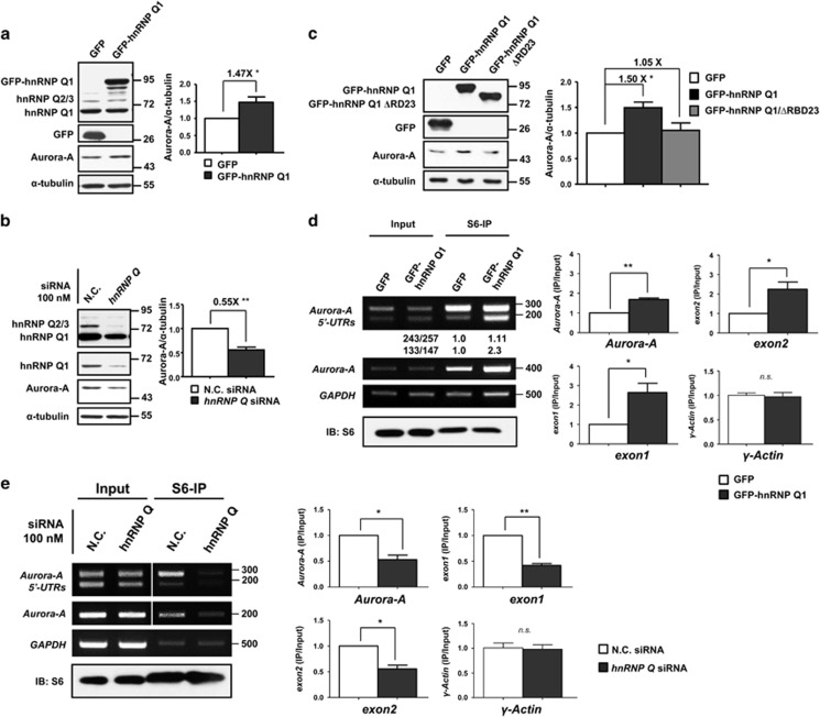
Figure 3.
HnRNP Q1 translationally upregulates Aurora-A mRNA and increases Aurora-A protein expression. (a and b) SW480 cells transiently transfected with GFP, GFP-hnRNP Q1 (a) or control siRNA (NC), hn RNP Q siRNA (b) were collected to perform western blot analysis using antibodies as indicated. The quantitative results from three independent experiments of Aurora-A proteins expression level by western blot analysis are also shown; *P<0.05, and **P<0.01. (c) SW480 cells transiently transfected with GFP, GFP-hnRNP Q1 or GFP-hnRNP Q1/ΔRBD23 were collected for western blot analysis. The quantification of Aurora-A proteins expression from three independent experiments is shown; *P<0.05. (d) GFP or GFP-hnRNP Q1 expressed SW480 cells were collected for ribosomal protein S6-IP assay. The amount of Aurora-A mRNA or Aurora-A 5′-UTR isoforms in ribosomal complex was determined by RT-PCR (left) or RT-qPCR (right) from three independent experiments. The relative amount of exon1- (133/147) and exon2-containing (243/257) 5′-UTR of Aurora-A mRNAs immunoprecipitated by S6 protein were quantified by Scion Image as shown below. Upper: 243/257. Lower: 133/147. (e) SW480 cells were transfected with hnRNP Q siRNA for 48 h and then an S6-IP assay was performed as described above. The results of RT-PCR (left) and RT-qPCR (right) from three independent experiments are shown. Equal amount of immunoprecipitated S6 protein was verified by western blot. γ-Actin was used as negative control. *P<0.05, and **P<0.01; n.s., no significance

