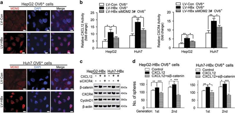
Figure 7.
HBx/MDM2-induced upregulation of CXCL12/CXCR4 activates the Wnt/β-catenin pathway in OV6+ liver CSCs. (a) Immunofluorescence assays were performed using the OV6+ HCC cells (HepG2 and Huh7) infected with either LV-HBx or LV-Con. The intracellular localization of MDM2 (red) was detected by confocal laser scanning microscopy as indicated. Nucleus was stained by DAPI dye (blue; scale bar=10 μm). (b) OV6+ LV-Con and LV-HBx HCC cells transfected with either negative control siRNA or siMDM2 were transfected with CXCL12 and CXCR4 luciferase reporters, followed by luciferase assay. The data display the mean±S.D., *P<0.05 and **P<0.01. (c) HBx-overexpressing HCC cells treated with control vehicle, recombinant CXCL12 protein alone or recombinant CXCL12 protein plus siRNA targeting CXCR4 for 48 h before collecting. The expression levels of indicated molecules in whole-cell lysates were detected by western blotting. β-actin was used as internal loading control. (d) The spheroid assay was performed to assess the number of spheres of HBx-expressing OV6+ CSCs treated with control vehicle, recombinant CXCL12 protein alone or recombinant CXCL12 protein plus siRNA targeting β-catenin. Experiments were performed in triplicate and all data represent the mean±S.D., *P<0.05, **P<0.01 and ***P<0.001

