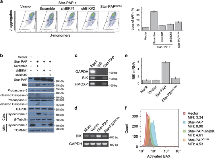
Figure 3.
Star-PAP induces mitochondrial apoptosis through regulating expression of BIK. (a) Loss of ΔΨm in SUM-159PT cells was measured by flow cytometry using JC-1 dye after being transfected with pCDNA3.1-Star-PAP and BIK shRNA/scramble, or Star-PAPD218A (left panel). Data from triplicate experiments were presented as mean±S.D. (right panel). (b) Cell lysates from SUM-159PT cells transfected with indicated plasmids were analyzed by western blot. For analyzing cytochrome c, mitochondrial and cytosolic fractions were separated. GAPDH was used as the loading control for whole cell lysates. β-Tubulin and TOMM20 were used as the cytosolic and mitochondrial loading control, respectively. (c) BIK mRNA was detected by RT-PCR after RIP of Star-PAP in SUM-159PT cells. GAPDH and HMOX-1 were used as the negative and positive control, respectively. (d) SUM-159PT cells were transfected with pCDNA3.1-Star-PAP or Star-PAPD218A, and BIK mRNA was examined by RT-PCR 24 h later. GADPH was used as the loading control. (e) After ectopic expression of Star-PAP or Star-PAPD218A in SUM-159PT cells, change of BIK mRNA level was quantified by qPCR and normalized to mock; n=3, error bar denotes S.D. (f) Activation of BAX was measured by flow cytometry using the conformation-specific antibody after being transfected with the indicated plasmids. Mean of MFI (mean fluorescence intensity) from two independent experiments is shown

