Figure 1.
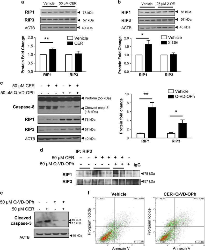
CER stimulates the necrosome in JEG3 cells. (a) Representative western blots and densitometric analysis of RIP1 and RIP3 kinases in choriocarcinoma JEG3 cells exposed to 50 μM C16:0 CER for 24 h. RIP3 protein levels were normalized to β-actin (ACTB) and expressed as fold change relative to vehicle control (EtOH). (b) Representative western blots and densitometric analysis for RIP1 and RIP3 kinases after exposure of JEG3 cells to 25 μM of 2-OE for 24 h. Values were normalized to ACTB and expressed as a fold change relative to vehicle control (DMSO). (a and b) Statistical significance was determined as *P<0.05 using an unpaired Student's t-test (n=5 different experiments carried out in triplicate). (c) Representative western blots for caspase-8, RIP1 and RIP3 following exposure of JEG3 cells to the pan-caspase inhibitor Q-VD-OPh (50 μM). Densitometric quantification of RIP1 and RIP3 levels following Q-VD-OPh treatment normalized to ACTB and expressed as a fold change relative to DMSO vehicle control. Statistical significance was determined as *P<0.05 or **P<0.01 using an unpaired Student's t-test (n=4 separate experiments, carried out in duplicate). (d) Immunoprecipitation (IP) of RIP3 followed by WB for RIP1/RIP3 in JEG3 cells treated with 50 μM CER alone or CER+Q-VD-OPh relative to vehicle control or 50 μM Q-VD-OPh alone (n=3 separate experiments). Goat IgG was used as a negative control. (e) Representative western blot of cleaved caspase-3 following exposure of JEG3 cells to 50 μM CER±50 μM Q-VD-OPh (n=3). ACTB was used as a loading control. (f) JEG3 cells treated with CER+Q-VD-OPh analyzed by flow cytometry Annexin V and propidium iodide double-labeling display an increase in necrotic cells (propidium iodide+) accompanied by a reduction of early apoptotic cells (Annexin V+/propidiumiodide−) compared with vehicle control cells. (n=3)

