Figure 4.
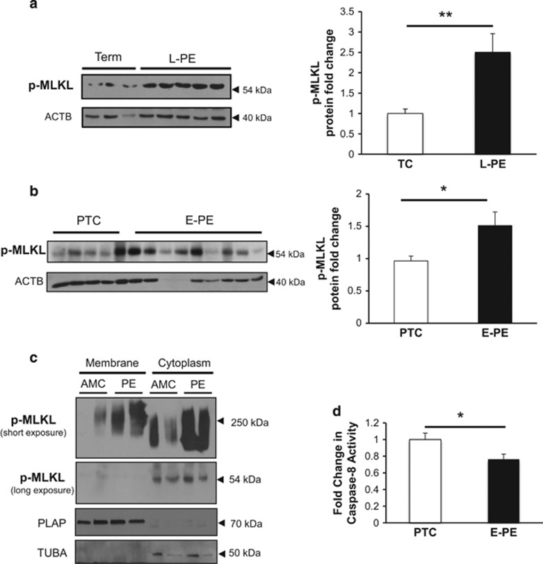
MLKL activation in preeclampsia associates with reduced caspase-8 activity. (a and b) p-MLKL protein expression in L-PE and E-PE placentae relative to TC and PTC controls as detected by western blotting. Densitometric analysis of p-MLKL expression in L-PE and E-PE placentae normalized to ACTB (L-PE, n=22; TC, n=10; E-PE, n=20; PTC, n=10). Statistical significance was determined as **P<0.01 using an unpaired Student's t-test. (c) Native PAGE analysis of membrane isolated from preeclamptic placentae and age-matched controls (AMC). Representative western blots demonstrated p-MLKL expression as an oligomer of high molecular weight (250 kDa) as well as a monomer of 54 kDa in the membrane and cytoplasmic fractions, respectively. PLAP represents the loading control for the membrane fractions, whereas TUBA was used as the cytoplasmic loading control. (d) Caspase-8 activity assay in placentae from E-PE and PTC (E-PE, n=6; PTC, n=6)

