Figure 7.
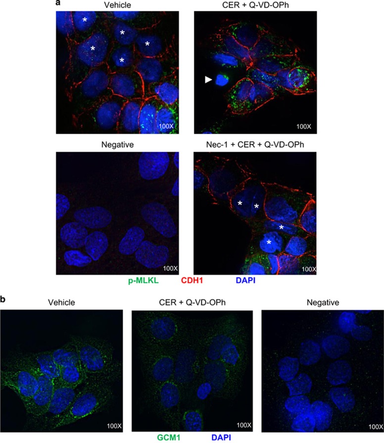
Necroptotic conditions impair trophoblast cell fusion. (a) Representative immunofluorescence images for p-MLKL (green) and CDH1 (red) in primary trophoblasts following exposure to 50 μM CER+Q-VD-OPh in the presence or absence of Nec-1. Asterisks designate fusing cell nuclei. Twenty-four-hour exposure to 50 μM CER+Q-VD-OPh led to increased expression of p-MLKL and retention of cell boundaries in primary trophoblast cells. A dying cell with shrunken nuclei is indicated by a white arrow and exhibits strong expression of p-MLKL punctae. Nuclei were counterstained with DAPI (blue). Rabbit and mouse IgG were used for the negative control. (b) Representative immunofluorescence images for GCM1 (green) in primary trophoblast cells subjected to 50 μM CER+Q-VD-OPh treatment. CER+Q-VD-OPh led to a reduction in GCM1 expression compared with vehicle control. Nuclei were counterstained with DAPI (blue). Rabbit IgG was used as a negative control. Images shown at × 100 magnification

