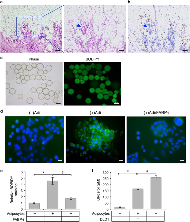
Figure 1.
Interaction of human colon cancer cells and cancer-associated adipocytes. (a) Abundant adipose tissues are closely associated with invasive colon cancers. H&E staining of colon cancer specimens collected from a patient diagnosed with stage IV colon adenocarcinoma. The enlarged image of the boxed region is shown on the right. The arrowhead indicates invasive tumor cells surrounded by the adipose tissue. Scale bar, 500 μm (left image) and 200 μm (right image). (b) Detection of infiltrated macrophages associated with tumor cells and the adipose tissue by IHC staining using the anti-CD68 antibody. Scale bar, 200 μm. (c) Phase contrast image and BODIPY-493/505 staining of human mature adipocytes isolated from colon cancer-associated adipose tissues. Scale bar, 100 μm. (d) SW480 cells were cultured alone or with human mature adipocytes for 48 h. FABP4 inhibitor (FABP-i) was included in one set of cells co-cultured with adipocytes. Lipid droplets in cells were detected using BODIPY-493/505 staining (green) and cell nuclei were stained with DAPI (blue). The co-culture experiments were performed in triplicates and repeated three times using adipocytes isolated from three different patients. Scale bar, 20 μm. (e) Representative images as shown in d were analyzed to determine the intensity of BODIPY-493/505 staining in SW480 cells using Nikon Elements AR software. Six randomly chosen images for each condition were quantified and data represent the mean±S.D. (*P<0.001). (f) DLD1 cells and primary human adipocytes were cultured alone or together in serum-free DMEM medium for 24 h. Lipolysis was detected by measuring the concentration of glycerol in the medium. Data represent the mean±S.D. (*P<0.001 and #P<0.01)

