Figure 2.
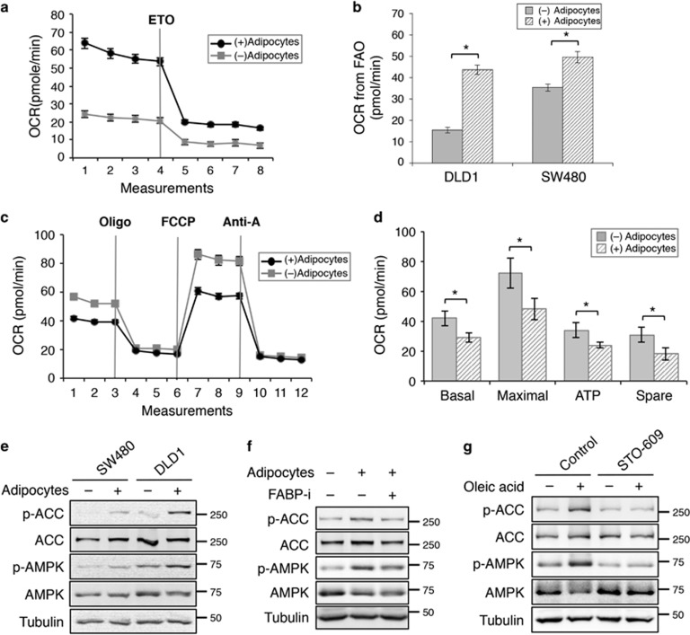
Uptake of fatty acids from adipocytes alters cellular metabolism in colon cancer cells. (a) Representative measurements of OCR in DLD1 cells after co-cultured with or without adipocytes using the Seahorse Bioscience XF96 Extracellular Flux Analyzer. The levels of OCR were decreased after FAO was inhibited after ETO treatment. (b) The amount of OCR derived from FAO was quantified as response to ETO treatment. Data represent the mean±S.D. (*P<0.001). (c) Representative measurements of mitochondrial function in SW480 cells after co-culturing with or without adipocytes for 24 h using the Seahorse Bioscience XF96 Extracellular Flux Analyzer. The cells were subjected to Mito Stress Tests in medium containing glucose (25 mM) and glutamine (2 mM). (d) The relative levels of OCR associated with basal respiration, maximal respiration, ATP-linked respiration, and spare respiratory capacity were calculated based on the measurements obtained upon the addition of different compounds. Data represent the mean±S.D. (*P<0.001). The OCR measurements were normalized to the protein contents. (e) SW480 and DLD1 were co-cultured with or without adipocytes for 48 h. Cell lysates were prepared and analyzed for phosphorylation and total protein expression of AMPK and ACC using western blotting. (f) Cell lysates were prepared from SW480 cells that have been treated with adipocytes in the presence or absence of FABP4 inhibitor and analyzed for phosphorylation and total protein expression of AMPK and ACC using western blotting. (g) Fatty acids activate AMPK directly. SW480 cells were treated with oleic acid (100 μg/ml) in the presence or absence of CaMKK2 inhibitor STO-609 (10 μM) for 24 h. Cell lysates were analyzed activation of AMPK and ACC using western blotting

