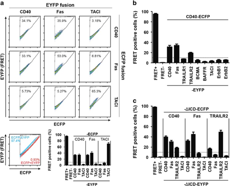
Figure 1.
CD40 interacts with Fas and TRAILR2. (a) Flow cytometry FRET assay between CD40, Fas and TACI. The image shows the combination of the three receptors fused to ECFP and EYFP. The gate used to calculate the percentage of FRET-positive cells was established by using a ECFP-EYFP fusion protein (100% FRET blue dots) together with a ECFP/EYFP co-transfection (0% FRET red dots), bottom left panel. Bottom right panel shows the mean value of FRET-positive cells and SEM of five independent experiments. (b) Flow cytometry FRET screening for different TNFRSF members expressed in B cells. (c) Flow cytometry FRET assay between CD40, Fas, TRAILR2 and TACI lacking the intracellular domain (ΔICD). In all cases, FRET+ corresponds to the positive FRET reporter (ECFP–EYFP fusion protein) and FRET- corresponds to the negative control (ECFP/EYFP co-transfection). The dotted line represents background FRET levels

