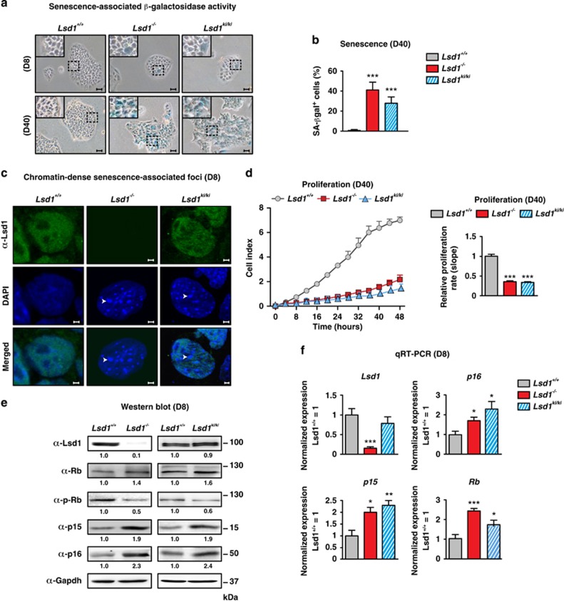
Figure 1.
Lsd1 deficiency boosts senescence in TSCs. (a–f) Comparison of wild-type (Lsd1+/+), Lsd1-deficient (Lsd1−/−) and Lsd1 enzymatic inactive mutant (Lsd1ki/ki) TSCs. (a and b) Representative images of senescent-associated β-galactosidase activity (blue) in TSCs (a) and quantification of senescent cells (b). (c) DNA stain by DAPI depicts chromatin-dense senescent-associated foci (arrows). TSCs were co-stained with an Lsd1 antibody. (d) Proliferation of TSCs was determined in real time. (e) Western blot decorated with the indicated antibodies including phosphorylated Rb (p-Rb). Band intensity was normalized to Gapdh as loading control relative to Lsd1+/+. (f) qRT-PCR of Lsd1 and indicated cell cycle regulators normalized to three housekeeping genes relative to transcription in wild-type TSCs. Scale bars: 10 μm (a) and 2 μm (c). Data were analyzed from at least three biological samples and are represented as mean+S.E.M. *P<0.05; **P<0.01; ***P<0.001 (unpaired, two-tailed Student's t-test, (b, d and f))

