Figure 5.
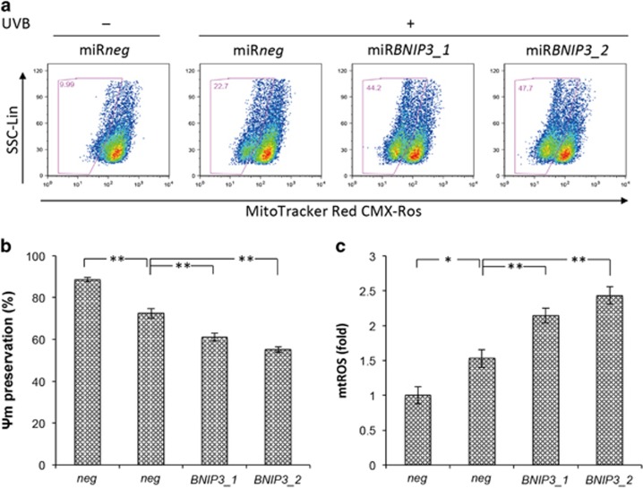
BNIP3 is required for the removal of dysfunctional mitochondria in epidermal keratinocytes after UVB exposure. HPEKs were infected with adenoviral vectors expressing shRNA followed by UVB radiation (20 mJ/cm2). (a and b) Mitochondrial membrane potential (ΔΨm) was measured using MitoTracker Red 4 h after UVB exposure. (a) Representative dot plots by flow cytometric analysis are shown. Gates represent cells with decreased ΔΨm. (b) Changes in ΔΨm were plotted as the means±S.E. of five independent experiments. **P<0.01. *P<0.05. (c) Mitochondrial ROS (mtROS) were measured using MitoSox Red. Graph indicates the quantification of mean fluorescence intensity (MFI) for MitoSox Red staining. Data are presented as the mean±S.E. from five independent experiments

