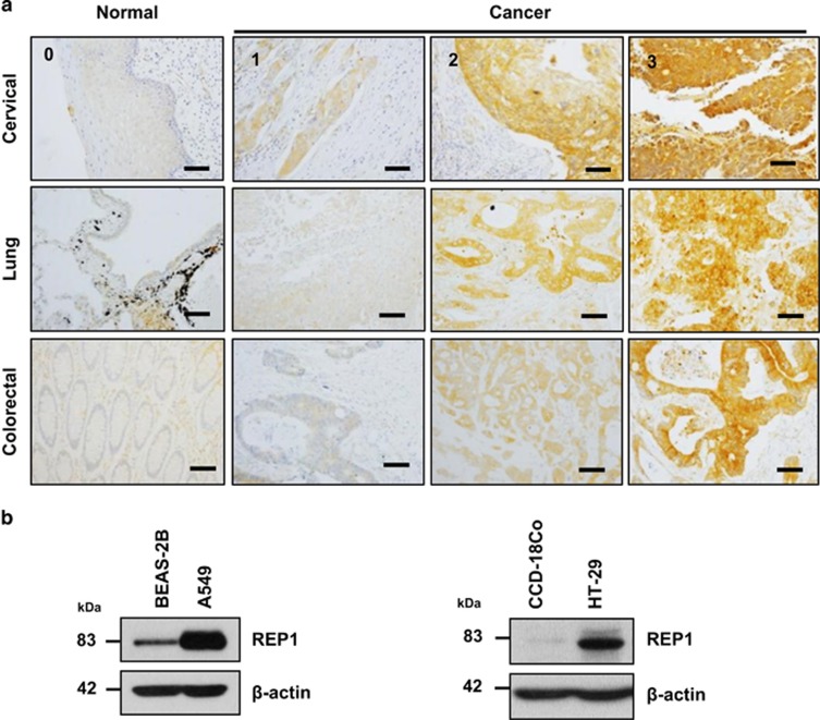
Figure 1.
REP1 expression in human cancer tissues and cancer cell lines. (a) Cancer patient-derived microarrays for cervical, lung, and colorectal tissue were examined for REP1 expression using an immunoperoxidase method. Staining results were graded according to the intensity and proportion of positive cells as described in ‘Materials and Methods'. Scale bar=50 μm. (b) A human normal lung epithelial cell line BEAS-2B and a lung adenocarcinoma cell line A549, and a normal colon epithelial cell line CCD-18Co and a colorectal adenocarcinoma cell line HT-29 were processed for immunoblot analysis using anti-REP1 antibody and β-actin antibody was used as a loading control. These experiments were performed three independent times with comparable results

