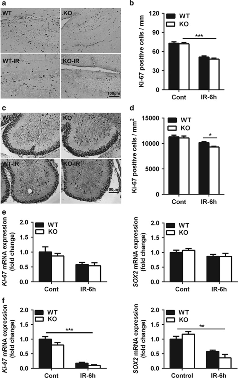
Figure 2.
Neural stem and progenitor cell proliferation in the dentate gyrus and cerebellum. (a) Representative Ki-67 immunostaining in the dentate gyrus. (b) Quantification of Ki-67-positive cells in the SGZ of the dentate gyrus showed a significant decrease after irradiation. (c) Representative Ki-67 immunostaining in a cerebellar lobule. (d) Quantification of Ki-67-positive cells in the EGL of a cerebellar lobule. (e) The mRNA expression of Ki-67 and SOX2 in the hippocampus. (f) The mRNA expression of Ki-67 and SOX2 in the cerebellum decreased significantly after irradiation; n=7 per group for the immunostaining; n=5 per group for the quantitative PCR (qPCR) assays. *P<0.05 and **P<0.01

