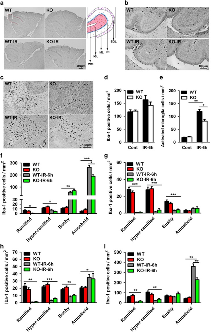
Figure 4.
Neuronal Atg7 deficiency reduces microglia activation in the cerebellum. (a) Representative Iba-1 immunostaining in sagittal sections of the cerebellum. Each folia comprises distinct cellular layers: EGL; ML; Purkinje cell layer (PC), IGL, and WM. (b) Representative Iba-1 immunostaining in the EGL, ML, and IGL of a cerebellar lobule. (c) Representative Iba-1 immunostaining in the WM of a cerebellar lobule. (d) Quantification of total Iba-1-positive cells in the whole cerebellum. (e) Quantification of total activated microglia based on morphology in the whole cerebellum. (f) Quantification of Iba-1-positive cells according to morphology in the EGL of the whole cerebellum. (g) Quantification of Iba-1-positive cells according to morphology in the ML. (h) Quantification of Iba-1-positive cells in the cerebellar IGL. (i) Quantification of Iba-1-positive cells in the WM of the whole cerebellum; n=7 per group. *P<0.05, **P< 0.01, and ***P<0.001

