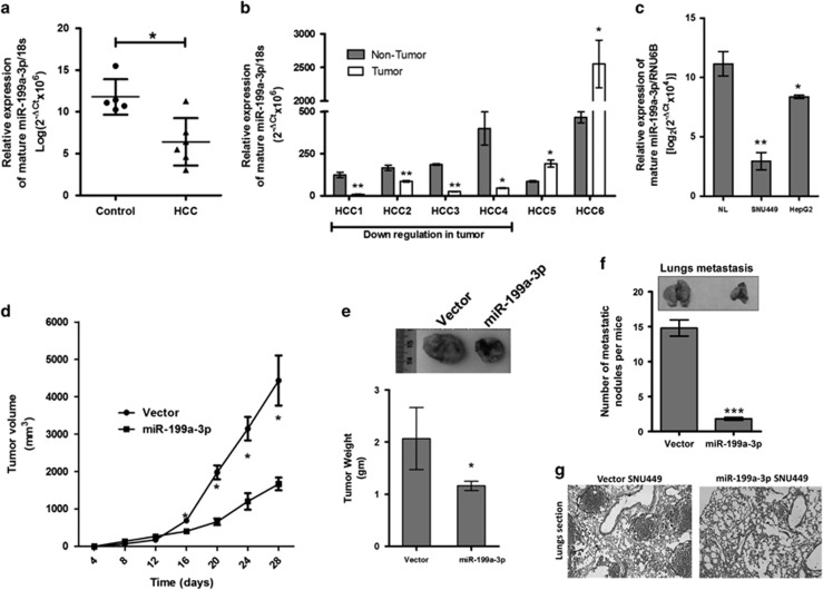
Figure 1.
Anti-tumorigenic role of miR-199a-3p. miR-199a-3p expression was compared between (a) HCC tumor and normal liver (NL) tissues, (b) HCC tumor and adjacent non-tumor tissues and (c) HCC cell lines and normal liver (NL). (d) Tumor volume at different time points and (e) tumor weight of subcutaneous HCC in either vector or miR-199a-3p overexpressed cell line injected NOD/SCID mice. (f) Size and number of the nodule formed in the lung after 4 weeks of injection of either vector or miR-199a-3p overexpressed cell line through tail vein in NOD/SCID mice. (g) Hematoxylin and eosin staining of the lung section. Area enclosed by dotted line indicates metastatic growth. *P<0.05, **P<0.01 and ***P<0.005 respectively

