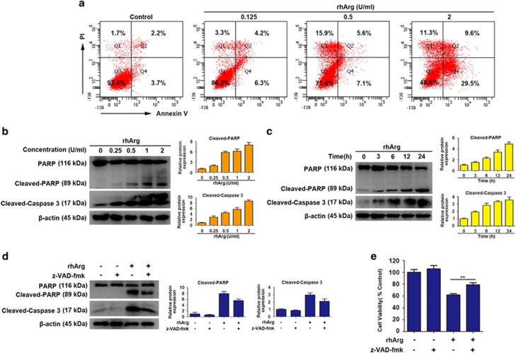
Figure 2.
RhArg induced caspase-dependent apoptosis in H1975 cells. (a) After treatment with different concentrations of rhArg for 72 h, H1975 cells were stained with Annexin V/PI and analyzed by flow cytometry. (b) H1975 cells were dose dependently treated with rhArg for 24 h. (c) H1975 cells were time dependently treated with 2 U/ml of rhArg. (d) H1975 cells were incubated with 2 U/ml of rhArg in the presence or absence of 20 μM z-VAD-fmk for 24 h. (b–d) Western blot analysis was performed to assess the expression level of PARP, cleaved-PARP and cleaved-caspase 3. Densitometric values were quantified using the ImageJ software and normalized to control. The values of control were set to 1. The data are presented as means±S.D. of three independent experiments. (e) H1975 cells were incubated with 2 U/ml of rhArg in the presence or absence of 20 μM z-VAD-fmk for 48 h. Results were expressed as mean±S.D. and analyzed by Student's t-test (two-tailed). **P<0.01

