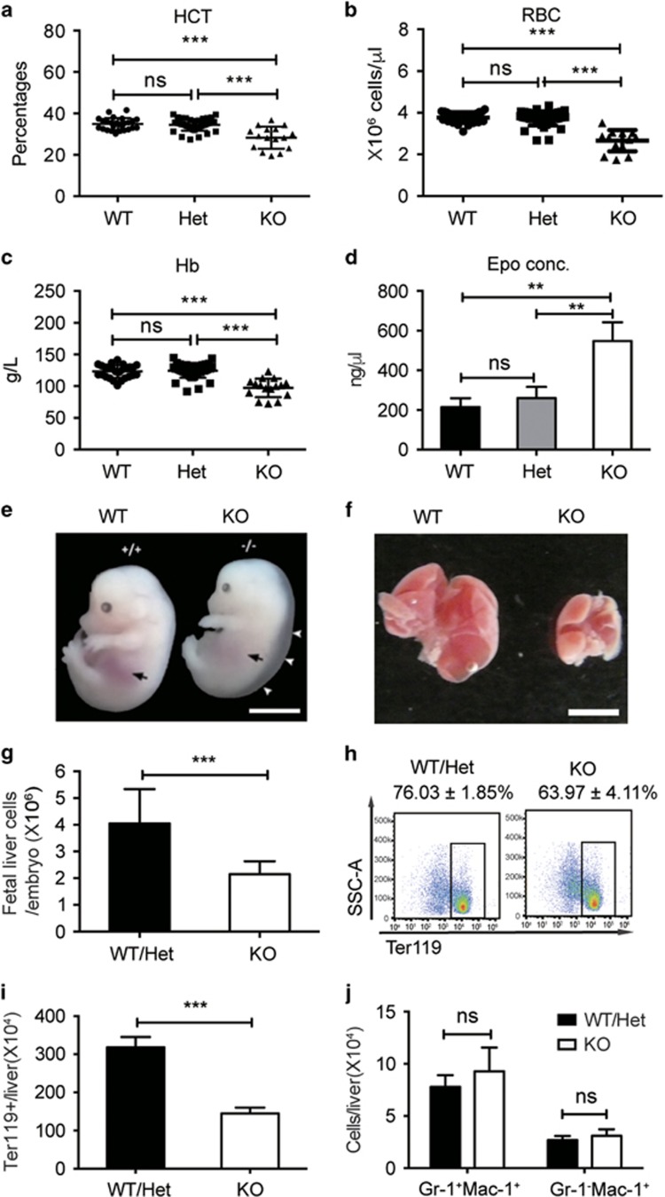
Figure 1.
Stk40 KO embryos have anemia. (a–c) Peripheral blood routine tests for E18.5 Stk40 WT, Het and KO embryos. HCT, hematocrit; RBC, red blood cell; Hb, hemoglobin. WT, n=26; Het, n=48; KO, n=17. ***P≤0.001; ns, no significance. (d) Concentrations of erythropoietin (Epo) from Stk40 WT, Het and KO embryos at E18.5. WT, n=6; Het, n=6; KO, n=6. **P≤0.01; ns, no significance. (e) Gross morphology of WT and Stk40 KO embryos at E14.5. White arrowheads indicate subcutaneous edema. Black arrows indicate the position of the fetal liver. Scale bars, 5 mm. (f) Gross morphology of representative fetal livers from Stk40 WT and KO embryos at E14.5. Scale bars, 2 mm. (g) Total number of fetal liver cells from Stk40 WT/Het and KO embryos at E14.5. (h) The representative frequencies of Ter119+ cells from Stk40 WT/Het and KO embryos at E14.5. (i) Absolute numbers of Ter119+ cells from Stk40 WT/Het and KO embryos at E14.5. (j) Absolute numbers of monocytes (Gr-1−Mac-1+) and granulocytes (Gr-1+Mac-1+) from Stk40 WT/Het and KO embryos at E14.5. For panels (g–j): WT, n=8; Het, n=30; KO, n=14. ***P≤0.001; ns, no significance

