Figure 6.
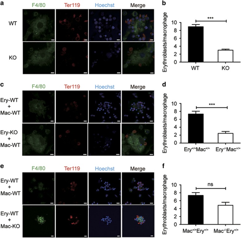
Stk40 is crucial for the EBIs formation. (a) Representative images of native EBIs isolated from fetal livers of E14.5 WT and Stk40 KO embryos. Islands were stained with F4/80-FITC (green) for macrophages, Ter119-APC (red) for erythroblasts, and Hoechst 33342 for the nuclei. Scale bar, 5 μm. (b) Numbers of erythroblasts bound per macrophage of E14.5 WT and Stk40 KO embryos. Sixty islands from nine embryos were counted for each genotype. ***P≤0.001. (c) Representative images of reconstituted islands from WT and Stk40 KO erythroblasts (Ery) incubated with WT macrophages (Mac) of E14.5 WT and Stk40 KO embryos. (d) Numbers of erythroblasts bound per macrophage. For each cell combination, total 40–60 islands from 10 embryos were counted. ***P≤0.001. (e) Representative images of reconstituted EBIs from WT erythroblasts (Ery) incubated with WT and Stk40 KO macrophages (Mac), respectively. (f) Numbers of erythroblasts bound per macrophage. For each cell combination, total 40–60 islands from 10 embryos were counted. Ns, no significance. For panels (c and e), scale bar, 10 μm

