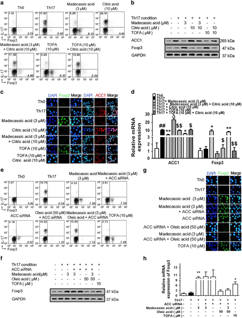
Figure 4.
Madecassic acid facilitated the shift of T helper type 17 (Th17) toward regulatory T cells by inhibiting ACC1 expression. (a) Naive T cells were cultured with madecassic acid (3 μM) and ACC1 activator citric acid (10 μM) under Th17-inducing conditions for 4 days. The proportions of CD4+IL-17+ T cells and CD4+Foxp3+ T cells were detected by flow cytometry. (b–d) Naive T cells were cultured with madecassic acid (3 μM) and ACC1 activator citric acid (10 μM) under Th17-inducing conditions for 4 days. The mRNA and protein expression of Foxp3 and ACC1 was analyzed by western blot, immunofluorescence and real-time PCR. (e) Naive T cells were cultured with madecassic acid (3 μM), oleic acid (50 μM) and ACC1 siRNA under Th17-polarizing conditions for 4 days. The proportions of CD4+IL-17+ T cells and CD4+Foxp3+ T cells were detected by flow cytometry. (f–h) Naive T cells were cultured with madecassic acid (3 μM), oleic acid (50 μM) and ACC1 siRNA under Th17-polarizing conditions for 4 days. The relative expression of Foxp3 was analyzed by western blot, immunofluorescence and real-time PCR. GAPDH was used as a cytoplasm marker. The data were expressed as means±S.E.M., n=3. #P<0.05, ##P<0.01 versus Th0 group; *P<0.05, **P<0.01 versus Th17 group; ΔP<0.05 versus Th17 group; $P<0.05, $$P<0.01 versus citric acid group

