Figure 5.
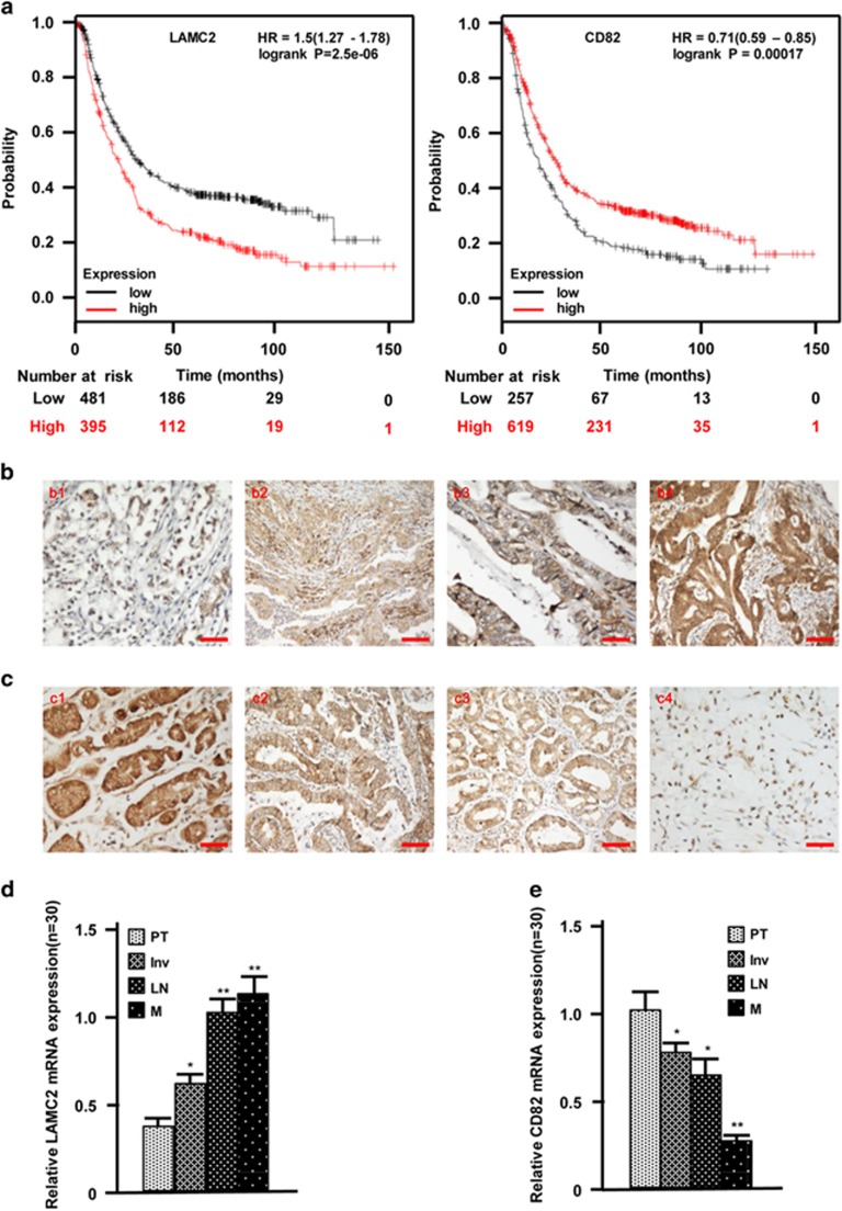
The enhanced LAMC2 and decreased CD82 indicate tumor metastasis and worse outcome of GC. (a) The overall survival of GC patients was evaluated by Kaplan–Meier plots. (b) The gradual increase of LAMC2 proteins were examined by IHC staining in GC specimens from the tumor in situ (b1), invasive tumor tissues (b2), tumors with lymph node metastasis (b3) and metastasis tissues (b4). (c) IHC staining to show the gradual decrease of CD82 proteins in GC tissues derived from the tumor in situ (c1), invasive tumors (c2), tumors with lymph node metastasis (c3) and tumors with distant metastasis locus (c4). (d and e) The expressions of LAMC2 (d) and CD82 (e) in various gastric carcinomas were quantitatively determined by qRT-PCR. PT: tumor in situ; Inv: invasive tumor; LN: tumor with lymph node metastasis; M: tumor with distant metastasis locus (*P<0.05, *P<0.01, versus PT). Scale bars, 100 μm

