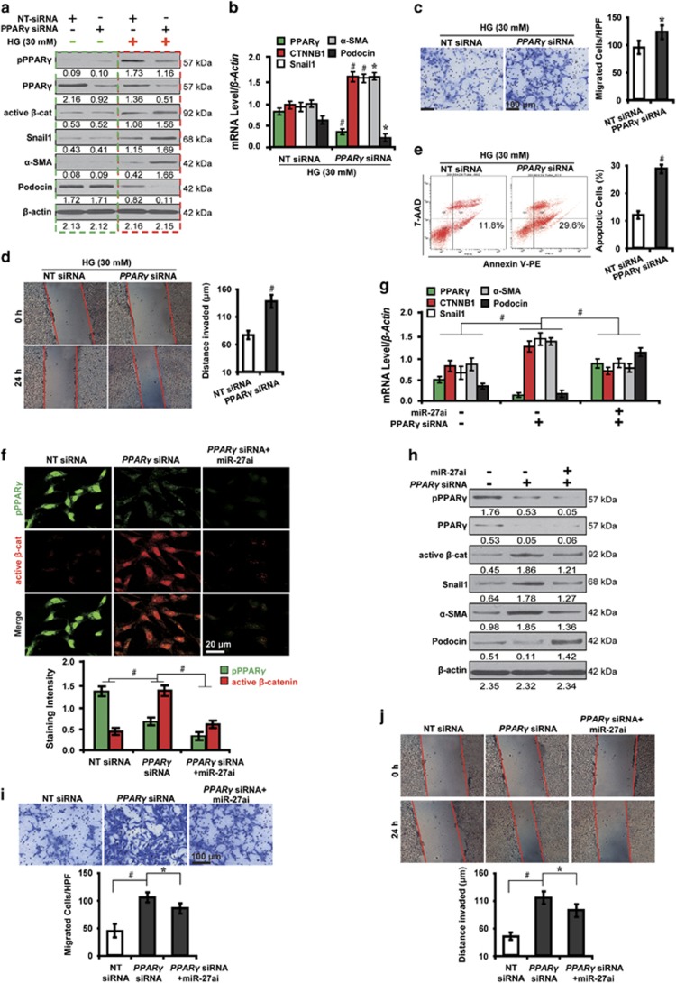
Figure 3.
PPARγ-mediated β-catenin activation induces podocyte injury in high glucose. (a) Representative western blotting shows the expression of phosphorylated and total PPARγ and β-catenin target genes in various conditions as indicated. (b) qRT-PCR analysis shows PPARγ siRNA decreased PPARγ and podocin but increased β-catenin target genes. (c) Transwell migration assay and quantitative data show increased migration. Scale bar, 100 μm. (d) Wound-healing assay and quantitative data show increased invasion. (e) Summarized data showing increased podocyte apoptosis by flow cytometric analysis. (f) Immunofluorescence microscopy and quantitative data show PPARγ siRNA-induced β-catenin activation was attenuated by co-transfection with miR-27ai. Scale bar, 20 μm. Representative (g) qRT-PCR and (h) western blotting show the expression of PPARγ and β-catenin target genes in various conditions as indicated. Representative (i) transwell migration assay and (j) wound-healing assay show the increased ability of migration and invasion caused by PPARγ abolishment were mitigated by co-transfection with miR-27ai. *P<0.05; #P<0.001. Active β-cat, active β-catenin; CTNNB1, catenin beta-1; NT, non-targeting; pPPARγ, phosphorylated peroxisome proliferator-activated receptor γ

