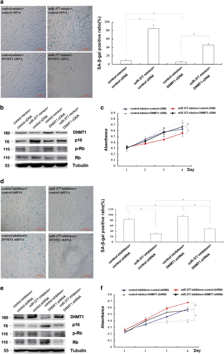
Figure 4.
miR-377 promoted senescence by suppressing DNMT1 expression in HSFs. (a) Cellular senescence was detected by evaluating SA-β-gal-positive cells in young HSFs (PD<10) treated with miR-377 mimics together with control cDNA or DNMT1 cDNA as indicated (left). The positive cell quantification was shown (right; data represent the mean±S.E.M. n=3, *P<0.05). (b) DNMT1, p16, and Rb expressions and phosphorylation level of Rb were detected by western blot in young HSFs (PD<10) treated with miR-377 mimics together with control cDNA or DNMT1 cDNA as indicated (*P<0.05). Representative data was shown. (c) Absorbance at 490 nm was detected by MTS assays in young HSFs (PD<10) treated with miR-377 mimics together with control cDNA or DNMT1 cDNA as indicated (data represented as the mean±S.E.M. n=3 at each time point, *P<0.05). (d) Cellular senescence was detected by evaluating SA-β-gal-positive cells in passage-aged HSFs (PD>50) treated with miR-377 inhibitors together with control shRNA or DNMT1 shRNA as indicated (left). The SA-β-gal-positive ratio was shown (right; data represented as the mean±S.E.M. n=3, *P<0.05). (e) DNMT1, p16, and Rb expressions and phosphorylation level of Rb were detected by western blot in passage-aged HSFs (PD>50) treated with miR-377 inhibitors together with control cDNA or DNMT1 cDNA as indicated (*P<0.05). Representative data was shown. (f) Absorbance at 490 nm was detected by MTS assays in passage-aged HSFs (PD>50) treated with miR-377 inhibitors together with control cDNA or DNMT1 cDNA as indicated (data represented as the mean±S.E.M. n=3 at each time point, *P<0.05)

