Figure 3.
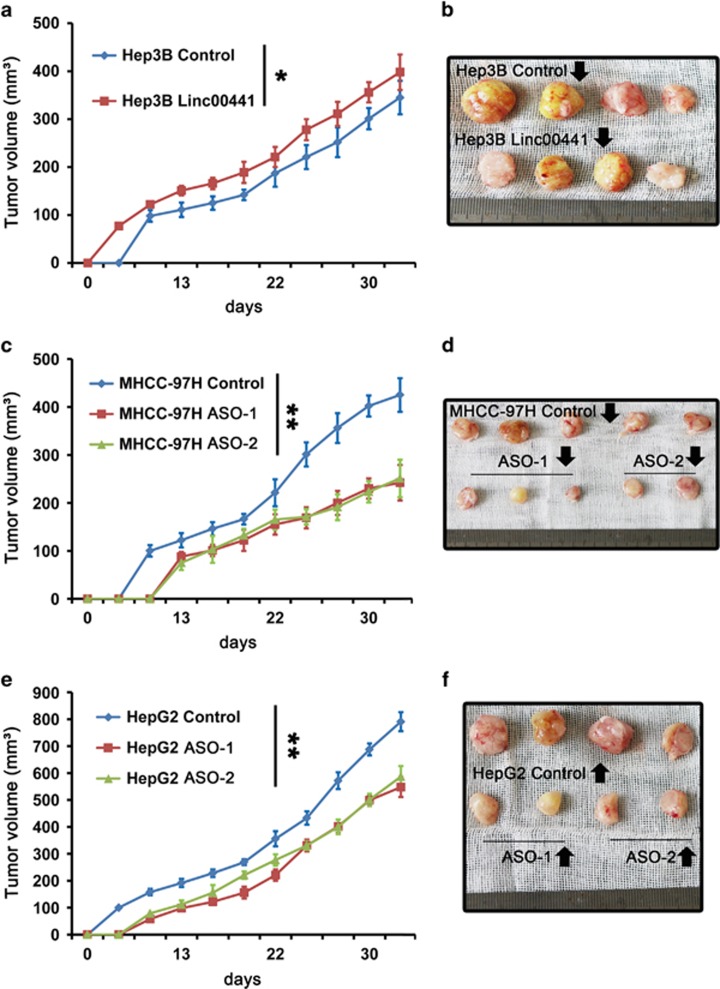
Linc00441 enhanced tumor growth in vivo. Mice with established tumors in different groups were measured approximately every 3 days and the tumors separated from killed mice at day 35 were presented in the right panel. (a and b) Cells treated with Linc00441 overexpression lentivirus. (c and d) Cells treated with Linc00441 ASOs and control group in MHCC-97H. (e and f) Cells treated with Linc00441 ASOs and control group in HepG2. Tumors were measured every week after the implantation, and the volume of each tumor was calculated (length × width2 × 0.5). Data are presented as means±S.E.M. (*P<0.05, **P<0.01)

