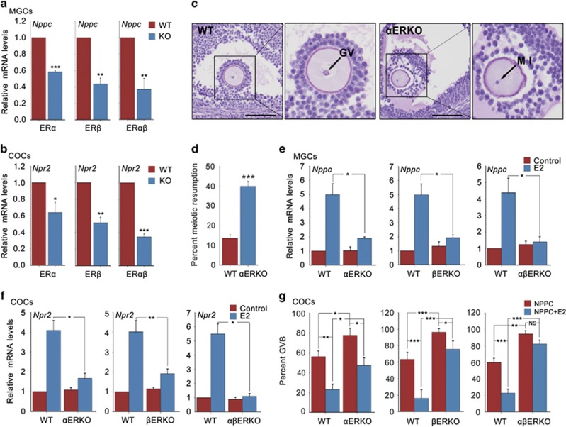
Figure 3.
ERKO mouse ovaries show decreased NPPC/NPR2 levels and percentages of meiotic arrested oocytes. (a and b) Expression of Nppc levels in MGCs and Npr2 levels in COCs isolated from 22- to 24-day-old WT and ERKO mouse ovaries. Ovaries were stimulated with PMSG for 46 to 48 h. n=3. (c) A prophase-arrested oocyte (GV) within a large antral follicle of a WT ovary and an oocyte with metaphase I (MI) chromosomes within a large antral follicle of a αERKO ovary. Scale bars: 100 μm. (d) Percentages of oocytes that had resumed meiosis, counted in serial sections of ovaries from WT and αERKO mice. n=9. (e and f) Effects of E2 on Nppc levels in MGCs and Npr2 levels in COCs isolated from WT and ERKO mice. MGCs and COCs were cultured for 24 h in medium without (control) or with 0.1 μM E2. n=3. (g) Effect of E2 on NPPC-maintained oocyte meiotic arrest within COCs isolated from WT and ERKO mice. COCs were incubated in medium containing 0.03 μM NPPC or plus 0.1 μM E2 for 24 h. n=4. Data represent the mean±S.E.M. *P<0.05, **P<0.01 and ***P<0.001 (t-test)

