Figure 5.
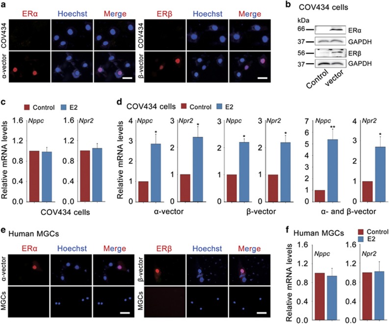
E2-ERs promote Nppc and Npr2 levels in human granulosa cells. (a) Expression of ERα and ERβ (red) in COV434 cells (upper) or COV434 cells after transfection with myc-tagged human α-vector or β-vector for 48 h (under). The nuclei were stained as blue by Hoechst. Scale bars: 25 μm. (b) WB analysis of ERα and ERβ protein levels in COV434 cells. COV434 cells were transfected with empty vector (control), myc-tagged human α-vector or β-vector for 48 h. GAPDH served as a loading control. (c) E2 failed to promote Nppc and Npr2 mRNA levels in COV434 cells. COV434 cells were cultured in medium without (control) or with 0.1 μM E2 for 24 h. Data represent the mean±S.E.M. n=3. (d) E2 increased Nppc and Npr2 mRNA levels in COV434 cells after transfection with myc-tagged human α-vector, β-vector or both. COV434 cells were incubated without (control) or with 0.1 μM E2 for 24 h. *P<0.05 and **P<0.01 (t-test). Data represent the mean±S.E.M. n=3. (e) Expression of ERα and ERβ (red) in human MGCs. Human MGCs were freshly isolated from ovulatory follicles, which were stimulated with FSH for 10 days, and followed by LH for 36 h. Human MGCs after transfection with myc-tagged human α-vector or β-vector for 48 h served as the corresponding positive control (upper). The nuclei were stained as blue by Hoechst. Scale bars: 25 μm. (f) Effect of E2 on Nppc and Npr2 mRNA levels in human MGCs. Human MGCs were freshly isolated from ovaries stimulated with FSH for 10 days followed by LH for 36 h and cultured for 24 h in medium containing 0.1 μM E2 or not (control). Data represent the mean±S.E.M. n=3

